Cervical stenosis


Cervical Stenosis: Definition
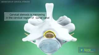
What is Cervical Stenosis?
Cervical Stenosis is a disorder in which the spinal canal in the neck area narrows. With less room in the spinal canal, the spinal cord may be compressed or pinched. Some people are born with this condition, but in most cases, it occurs due to the normal "wear and tear" of aging. Cervical Stenosis is most common in people over the age of 50.
Many people with Cervical Stenosis have suffered some type of trauma or injury to their neck, but their injury may have occurred months or years before they begin to develop symptoms of Cervical Stenosis.

Reasons
To understand what happens in Cervical Stenosis of the spine, it's necessary to understand the process of disc degeneration. This is a "wear and tear" process. To help you get a better idea of what happens in your spine, think about two vanilla wafers with a marshmallow between them. The wafers represent two vertebrae and the marshmallow represents an intervertebral disc. If you squeeze the wafers, the marshmallow has some "give" because it's fresh and pliable. It's soft like a pillow. If you leave the marshmallow out on the counter for a week, it will dry out. If you put it between the wafers after it has dried out, it isn't soft and won't provide the cushion for the wafers like it did when it was fresh. It may even split or tear. If you left the marshmallow out for a year, it would be so shrivelled and dry, it wouldn't be any good as a "shock absorber" at all. This is similar to what can happen in Cervical Stenosis.
As the body ages, and Cervical Stenosis occurs, the intervertebral discs lose some of their water content. This means they become dehydrated and lose some of their ability to act as shock absorbers and tears may occur in the outer ring of the discs. These tears in the outer ring may not cause any symptoms, so the person may not be aware an injury leading to Cervical Stenosis has occurred. These tears heal by laying down scar tissue which is not as strong as normal disc tissue. As more tears occur, more scar tissue is laid down. More wear and tear injuries keep occurring as time goes on. As the disc ages, it becomes less of a cushion and eventually cannot function as an effective shock absorber.
Eventually, the disc starts to collapse and the space between the vertebrae shrinks. When the disc collapses, this also impacts the way the vertebrae line up with each other. When the bones don't line up like they are meant to, there is less room in the spinal canal which leads to Cervical Stenosis and abnormal pressure is placed on joint surfaces, on the articular cartilage. The articular cartilage is the smooth covering over the end of bones where they come together in a joint. As time goes by, the abnormal pressure causes inflammation and arthritis to develop in the joints. Arthritis also leads to Cervical Stenosis.
Bone spurs may also start to form around the joints and the disc. It could be that too much movement in part of the spine could cause the formation of bone spurs. Eventually, bone spurs may form around the spinal nerves and Cervical Stenosis is the result.
Prevalence
Many people with Cervical Stenosis even though it may be severe, do not know they have the condition. It is apparent on x-ray findings in 5% of all adults. Cervical Stenosis occurs in 7% of people who are at least 50 years old and in 9% of adults over the age of 70 years.
Cervical Stenosis is usually caused by age-related changes in the size and shape of the spinal canal. Everyone experiences some degeneration with aging. People aged 50 years or older are at an increased risk of developing degenerative arthritic changes. As a person ages, the vertebral disc loses some of its water content and, as a result, loses some of its shock absorbing ability. As the disc continues to wear down, it begins to collapse, making the space between each vertebra smaller. The collapse also affects the way that the facet joints (the joints at the back of the spine) line up. Over time, this wear and tear damage from Osteoarthritis of the spinal bones can prompt the formation of bone spurs. As the bone spurs form, the size of the spinal canal becomes smaller and soon may begin to press on the spinal cord or its nerve roots.
Risk Factors
Certain factors increase the risk of developing Cervical Stenosis. They include:
-
- - Being born with a spinal canal that is more narrow than normal
-
- - Being female increases the risk of Cervical Stenosis
-
- - Being older than 50 years
-
- - Having a history of a spinal injury or spinal surgery
-
- - Having a history of Osteoarthritis or bone spurs can increase the risk of Cervical Stenosis
-
- - Having a medical history of inflammatory spondyloarthritis (for example Ankylosing Spondylitis)
-
- - Having a history of spinal tumors can increase the risk of Cervical Stenosis
- - Having a history of Paget's disease
Complications
If Cervical Stenosis is left untreated, or on rare occasions, treated too late, the patient may have the following complications:
-
- - Paralysis
-
- - Permanent Weakness
-
- - Permanent Numbness
-
- - Balancing problems
- - Incontinence
All of these complications that may arise from untreated Cervical Stenosis can really be a burden to the patient. If symptoms of Cervical Stenosis are detected or diagnosed, treatment should be done immediately to prevent the aforementioned complications to occur.
Cervical Stenosis: Symptoms and Diagnostic Procedures
Cervical Stenosis without compression of the spinal cord usually produces Cervical Stenosis symptoms such as neck pain and symptoms that are related to compression of the nerve root. Cervical Stenosis symptoms can include changes in sensation or changes such as numbness, tingling and abnormal feelings in the arm. Sometimes these Cervical Stenosis symptoms overlap with symptoms of spinal cord involvement and can include the gradual onset of weakness or clumsiness and numbness in the fingers and hand.
Cervical Myelopathy
Myelopathy is dysfunction of the spinal cord. Cervical Stenosis is one condition that can cause myelopathy, but many other conditions can also cause dysfunction of the spinal cord. When the nerves and membrane outside of the spinal cord are injured, pain results. This occurs in conditions like disc disease, fractures, arthritis or tumors outside the spinal cord. When the spinal cord is injured, it responds by producing weakness in the extremities, spasticity or tightness, alterations in sensation and possible problems with bowel and bladder functions. Patients who have spinal cord injuries may lose their "position sense." This is the ability to "know" the location of your extremities, even when your eyes are closed.
An examination by a physician can confirm these findings and the doctor may also note that the reflexes are hyperactive as well.
It's vital to determine the cause of a spinal cord problem because if the problem is corrected early, spinal cord function may be restored either partially or fully. Prolonged compression of the spinal cord may result in permanent damage and disability.
Over time, the pressure on the spinal cord as it runs through the cervical spine can cause the following Cervical Stenosis symptoms:
-
- Neck pain or stiffness. This is one of the most common Cervical Stenosis symptoms. The neck may be sore with the limitation of movement. The pain may not always be severe. In some cases, the neck may make grinding sounds, called crepitus, with certain movements.
-
- Changes in coordination or fine motor skills of the arms. The person may encounter trouble with typing, handwriting, buttoning a shirt, or putting a key in a door.
-
- Weakness and spasticity in the legs. Spasticity means a person loses control over his legs and may have a great deal of difficulty walking due to loss of control of where he places his feet. Balance problems may occur if the legs are not going where intended, requiring more reliance on a walking cane or hand rails.
-
- Numbness, weakness, burning or tingling sensation, and pins and needles in both the upper extremities and lower extremities. Hand weakness can be severe enough to affect a person’s grip. Intermittent shooting pains resembling an electric shock may extend the arms and legs, especially when bending the head forward (Lhermitte phenomenon).
-
- Changes in reflexes (may be increased in the legs) may be one of the cervical Cervical Stenosis.
-
- Loss of “position sense” which hinders a person to know where his arms and legs are when his eyes are closed.
- In severe cases, bowel or bladder dysfunction may also occur and can be one of the cervical Cervical Stenosis.
These Cervical Stenosis symptoms should be taken seriously and immediate consultation with a physician is warranted. If treatment is not sought, the spinal cord can become more compressed and more severe symptoms could develop, such as paralysis in one or more limbs or other bodily functions may shut down.
Diagnostic procedures
To diagnose Cervical Stenosis, the doctor will start by asking you how your Cervical Stenosis symptoms began and how they have progressed. After obtaining your medical history, your doctor will complete a physical examination that is focused mainly on your neck and the nerve function in your legs and arms. Your balance and your gait will most likely be tested if your doctor suspects Cervical Stenosis.
Your doctor may request X-rays to check for signs of Cervical Stenosis. These may show some signs of degeneration in the facet joints or disc spaces.
Magnetic resolution imaging: An MRI of your neck might be recommended if your doctor suspects Cervical Stenosis. These images enable your physician to see any structures that may be pinching or compressing the spinal cord or the nerves. Sometimes a dye is injected to allow further imaging and this is then followed by Computerized Tomography (CT scans).
Computed Tomography (CT) scan provides excellent detail of the bony tissues and some information about central soft-tissue abnormalities. The use of intravenous contrast agents improves the soft-tissue resolution significantly. The use of intrathecal contrast introduces some increased risk of injury, the risk of infection, and added expense. This should be limited to trauma cases and to those patients who have contraindications for MRI.
Electromyogram [EMG] and nerve conduction studies: These special studies can help your doctor distinguish if your pain is being caused by Cervical Stenosis or if it might be from other disorders such as Carpal Tunnel Syndrome. Some tests such as can study the conduction of signals through the spinal cord, and other tests study the signals sent to and from the nerves and muscles.
Cervical Spinal Stenosis Treatment
Non-Surgical Treatment
In some cases, the first cervical spinal stenosis treatment recommended is the immobilization of the neck. This can help decrease inflammation and pain. You may be instructed to limit your daily activities and to avoid all repetitive movements of the arms, neck and upper body. Your doctor may also want you to wear a soft neck brace for Cervical Stenosis. This brace is a padded ring that fits around the neck and attaches with a Velcro closure. It is usually worn during the day for up to three months, then the amount of time-worn is gradually decreased as your symptoms of Cervical Stenosis resolve.
Physical therapy is also sometimes recommended for Cervical Stenosis. The first cervical spinal stenosis treatment of therapy is aimed at pain control and relief of inflammation. Electrical stimulation therapy can help to relieve muscle spasms caused by cervical stenosis. Traction is sometimes used to gently stretch the muscles and joints. This can be accomplished by the therapist using a hand traction pull or with a special head halter designed for treating spinal conditions like Cervical Stenosis.
Epidural injections with cortisone medications are sometimes used in the cervical spinal stenosis treatment. These injections are administered into the portion of the spinal canal known as the epidural space. This space is located between the material that covers the spinal cord (the dura) and the spinal column. Steroids work to relieve inflammation around the discs and nerves. When the swelling is reduced, compression on the spinal cord is relieved and pain and inflammation of cervical stenosis may be decreased.
There are several surgical options for Cervical Stenosis, but they are typically divided into 2 categories. The anterior cervical decompression and fusion and the posterior cervical decompression.
-
- The anterior cervical decompression involves approaching the cervical spine from the front and removing any discs, bone spurs, or other structures that might be impinging the spinal cord. It typically includes fusing one or more levels of the cervical spine to maintain stability.
- The posterior cervical decompression involves approaching the cervical spine from the back, the bony arch at the back of the vertebra, called the lamina, is usually removed (laminectomy) or restructured (laminoplasty) to release pressure on the spinal cord. This procedure may or may not include a fusion.
Surgical Treatment
If conservative cervical spinal stenosis treatments are not successful in treating your Cervical Stenosis your doctor might recommend surgery. There are different types of procedures that can be used to treat the cervical stenosis. The goal of different surgical decompression techniques is to relieve pressure on the spinal cord by widening the spinal canal and removing the source of the compression. All surgical procedures come with a certain degree of risk, so your surgeon will discuss with you which option is most appropriate for your cervical stenosis.
- A decompressive laminectomy is commonly performed for cervical spinal stenosis treatment. In this procedure, the lamina or top section (roof) of the vertebra is removed. This creates more room in the spinal canal. If only a part of the lamina needs to be removed, the procedure is referred to as a laminotomy. A posterior laminoplasty may also be performed to help retain spinal stability in some cases of Cervical Stenosis.
- If you have been diagnosed with Cervical Stenosis, your surgeon may recommend a corpectomy and strut graft as a cervical spinal stenosis treatment. In this procedure, the surgeon removes the body of the vertebra and any bone spurs that are compressing the spinal cord. The vertebra that was removed is then replaced with a graft, which is a solid piece of bone, known as a strut graft. The graft heals over time and the spine fuses together where the vertebra was removed.
- If any intervertebral discs are herniated, they are removed to correct cervical stenosis in a procedure known as a discectomy. This increases room in the spinal canal and serves as a cervical spinal stenosis treatment. If the area where the nerve root exits the spinal canal (the foramen) needs to be enlarged to correct cervical stenosis, it is performed in a procedure called a foraminotomy.
- For patients who have Cervical Stenosis and a great deal of spinal instability, it may be necessary to perform spinal fusion in addition to decompression surgery. In spinal fusion surgery, a small piece of bone is taken from a donor site (usually the hip) and grafted into the spine. Surgical hardware, known as instrumentation, is used to help support the spine and may include plates, cages, rods, and screws. It can usually be determined prior to cervical spinal stenosis treatment surgery if fusion will be needed.
Care and Recovery
The main goal of a cervical spinal stenosis treatment will be rest. The patient may be able to walk with assistance within 24 hours after surgery. Activity is gradually increased over the course of the first 3 days. and patients are typically able to go home within a few days after the procedure, depending on the extent of the surgery. Adequate rest is still recommended once the patient is sent home. Some degree of pain and discomfort may be experienced during recovery. For this reason, patients are prescribed with oral analgesics. However, symptoms should begin to subside within a week or two after surgery. After two to four weeks, most patients can return to work or school and will no longer need their prescribed pain medications. Complete recovery typically occurs between two to six months after cervical spinal stenosis surgery.

 Spine
Spine